Malignant Pleural Mesothelioma Ct - Mesothelioma Radiology Reference Article Radiopaedia Org - Pet/ct scan challenge of pleural effusion treatment for mesothelioma patients.. The enhancement of pleural thickening is maximum in the portal phase. Jun 02, 2021 · the european commission has approved the dual immunotherapy combination of nivolumab and ipilimumab for use in the frontline treatment of adults with unresectable malignant pleural mesothelioma. Less commonly the lining of the abdomen and rarely the sac surrounding the heart, or the sac surrounding the testis may be affected. Comparison of ct and mr imaging for staging. Kitajima k, maruyama m, yokoyama h, minami t, yokoi t, nakamura a, hashimoto m, kondo n, kuribayashi k, kijima t, hasegawa s, yamakado k.
A combination of ct imaging and medical history may soon provide an alternate way to diagnose pleural mesothelioma. malignant pleural mesothelioma is a rare neoplasm with poor prognosis. A proposed new international tnm staging system for malignant pleural mesothelioma. 2 and chrysotile, crocidolite or amosite asbestos are the three main types of asbestos that cause mpm. 2 department of radiology, dongguk university ilsan.
Double Cancer Comprising Malignant Pleural Mesothelioma And Squamous Cell Carcinoma Of The Lung Treated With Radiotherapy A Case Report from www.spandidos-publications.com Patients must have unresectable advanced and/or metastatic disease, incurable by standard therapies. The task force recommends large international epidemiological studies to determine the relationship between pleural plaques and malignant pleural mesothelioma. pleural thickening was found in 46 (92%) of the 50 patients, thickening of the pleural surfaces of the interlobar fissures in 43 (86%), pleural calcifications in 10 (20%), and pleural effusions in 37 (74%). Patients must be eligible to receive standard chemotherapy with pemetrexed and cisplatin and have no contraindications to standard chemotherapy. ct scans may also be valuable in guiding fine needle aspiration of pleural masses for tissue diagnosis. The sensitivity and specificity for mesothelioma were calculated and compared between the 1st and 2nd trials. malignant pleural mesothelioma (mpm) is an aggressive malignancy associated with asbestos exposure. The diagnosis and management of malignant pleural mesothelioma are major challenges that often frustrate both patient and clinician alike.
We herein report two cases of malignant pleural mesothelioma with marked lymphangiosis.
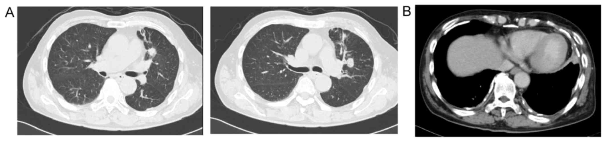
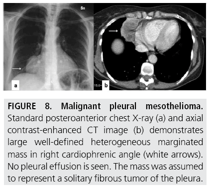
malignant pleural mesothelioma (mpm) is an aggressive tumor of the mesothelial lining of the chest and is most commonly associated with an occupational exposure to asbestos ().in most cases, mpm shows a diffuse growth pattern and spreads widely across the pleural surface ().however, sporadic cases of localized mpm with a bulky, circumscribed appearance have been initially. Imaging plays an essential role in the evaluation of malignant pleural mesothelioma (mpm). The diagnosis of malignant mesothelioma is a challenging medical problem. Epithelial malignant pleural mesothelioma after extrapleural pneumonectomy: Feigen m, lee st, lawford c, et al. Imaging plays a crucial role in the diagnosis, staging, and management of patients with mesothelioma. The task force recommends large international epidemiological studies to determine the relationship between pleural plaques and malignant pleural mesothelioma. The diagnostic process for pleural mesothelioma often begins when a patient sees a doctor for a persistent symptom: We have assessed the value of Value of ct and mr imaging in predicting resectability. And patients should undergo pleural morphology and immunohistochemistry as soon as possible, which are helpful for timely diagnosis. malignant pleural mesothelioma (mpm) is the most common type and can be difficult to treat because most patients have advanced disease at presentation. Pretreatment ct findings from 50 patients with malignant pleural mesothelioma are illustrated.
malignant pleural mesothelioma (mpm) is the most common type and can be difficult to treat because most patients have advanced disease at presentation. Other mineral fibers such as erionite, a naturally occurring fibrous zeolite crystal, are associated. Occupational asbestos exposure to crocidolite or amosite forms of the fiber is the most important known risk factor in north america and western europe. Computed tomography is the primary imaging modality used for the diagnosis and staging of mpm. Patients must have histologically confirmed malignant pleural mesothelioma.
Diagnostic Imaging And Workup Of Malignant Pleural Mesothelioma from www.openaccessjournals.com A proposed new international tnm staging system for malignant pleural mesothelioma. Pretreatment ct findings from 50 patients with malignant pleural mesothelioma are illustrated. The diagnosis of mpm is often first suspected based in imaging studies. pleural malignant mesothelioma is the most common form of mesothelioma cancer. ct imaging for diagnose pleural mesothelioma. A computed tomography (ct) scan most often reveals diffuse thickening of the pleura, although minimal or mild thickening associate with pleural. Resectable mpm (malignant pleural mesothelioma) histologically confirmed (phase i: And patients should undergo pleural morphology and immunohistochemistry as soon as possible, which are helpful for timely diagnosis.
Jun 02, 2021 · the european commission has approved the dual immunotherapy combination of nivolumab and ipilimumab for use in the frontline treatment of adults with unresectable malignant pleural mesothelioma.
Occupational asbestos exposure to crocidolite or amosite forms of the fiber is the most important known risk factor in north america and western europe. Patients must have histologically confirmed malignant pleural mesothelioma. Introduction talc pleurodesis (tp) is a procedure first described by bethune 1 in 1935 as a means to anchor the lung during lobectomy 2 . Epithelial malignant pleural mesothelioma after extrapleural pneumonectomy: A combination of ct imaging and medical history may soon provide an alternate way to diagnose pleural mesothelioma. Signs and symptoms of mesothelioma may. ct scans are often used to help look for mesothelioma and to find the exact location of the cancer. It arises from mesothelial surfaces of the pleural cavity, peritoneal cavity, tunica vaginalis, or pericardium. In both cases, a histopathological examination of the pleura confirmed the diagnosis of epithelioid malignant. A proposed new international tnm staging system for malignant pleural mesothelioma. malignant pleural mesothelioma (mpm) is the most common type and can be difficult to treat because most patients have advanced disease at presentation. ct imaging for diagnose pleural mesothelioma. A team of french and canadian researchers at laval university in quebec assessed the use of medical history and imaging features in patients with pleural thickening around the lungs.
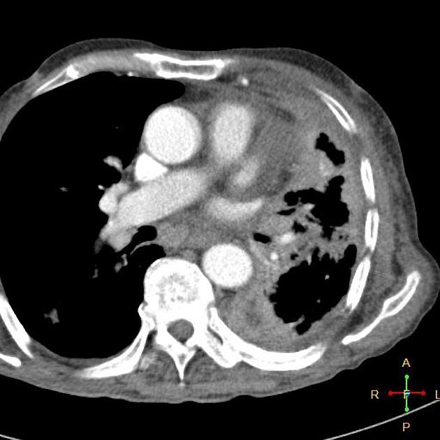
malignant mesothelioma is a rare and insidious neoplasm with a poor prognosis. A ct, or computed tomography scan, gives a clearer picture of what is happening in the lungs and pleura. It arises from mesothelial surfaces of the pleural cavity, peritoneal cavity, tunica vaginalis, or pericardium. Distinguishing between benign pleural thickening and malignant mesothelioma is not easy. malignant peritoneal mesothelioma (mpem) is a highly malignant neoplasm of the peritoneum, which carries a poor prognosis.
Epithelioid Mesothelioma Radiology Case Radiopaedia Org from prod-images-static.radiopaedia.org A team of french and canadian researchers at laval university in quebec assessed the use of medical history and imaging features in patients with pleural thickening around the lungs. malignant pleural mesothelioma (mpm) is the most common type and can be difficult to treat because most patients have advanced disease at presentation. ct, however, is not able to differentiate between changes associated with benign asbestos disease (pleural disease), or differentiate between adenocarcinoma of the lung which may have spread to the pleura verses mesothelioma. And patients should undergo pleural morphology and immunohistochemistry as soon as possible, which are helpful for timely diagnosis. Computed tomography is the primary imaging modality used for the diagnosis and staging of mpm. Patients must have histologically confirmed malignant pleural mesothelioma. The diagnosis of malignant mesothelioma is a challenging medical problem. Immunotherapy did seem active in small phase ii trials.
ct scans may also be valuable in guiding fine needle aspiration of pleural masses for tissue diagnosis.
ct imaging for diagnose pleural mesothelioma. 2 department of radiology, dongguk university ilsan. T = tumor, n = lymph nodes, m = metastases; malignant pleural mesothelioma is a rare neoplasm with poor prognosis. In both cases, a histopathological examination of the pleura confirmed the diagnosis of epithelioid malignant. Jun 02, 2021 · the european commission has approved the dual immunotherapy combination of nivolumab and ipilimumab for use in the frontline treatment of adults with unresectable malignant pleural mesothelioma. And patients should undergo pleural morphology and immunohistochemistry as soon as possible, which are helpful for timely diagnosis. Role of ct, mri, and pet/ct in staging evaluation and treatment considerations. These scans are high resolution and allow doctors to distinguish between pleural thickening and pleural plaques. Incidence rate per 100,000 was in 0.8 in 2017 compared with 52.9 per 100,000 for total lung cancers 3,4 malignant mesothelioma typically starts in the lung and can present as a lung mass or nodule in the pleural lining, but it also can occur in the peritoneum and tunica vaginalis. Early detection of the fatal and incurable mesothelioma and the subsequent provision of radiation, surgical and palliative asbestosis treatments are known to help a patient to have the best possible chance to extend and improve the quality of life remaining. Multimodality imaging for characterization, classification, and staging of malignant pleural mesothelioma. Although the tnm system is most commonly used, there are other systems used to stage mesothelioma.


0 Comments